Neck Muscle Diagram Front - Bim Lab Three Exercises To Improve Your Neck Posture Balance In Motion - The anterior and middle scalenes originate from the transverse processes of certain cervical.
byAdmin•
0
Neck Muscle Diagram Front - Bim Lab Three Exercises To Improve Your Neck Posture Balance In Motion - The anterior and middle scalenes originate from the transverse processes of certain cervical.. The infrahyoid muscles are ribbon like and contain following 4 matched muscles: The anterior and middle scalenes originate from the transverse processes of certain cervical. Attach the front panel module differs depending on the ca. Human muscle system, the muscles of the human body that work the skeletal system, that are under voluntary control, and that are it is accomplished primarily by the sternocleidomastoid muscles, with assistance from the longus colli and the longus capitis, which are found in the front of the neck. The muscular anatomy of the neck is correlated chiefly with movements of the head and shoulders.
Sternocleidomastoid muscle (main muscle in the front of the neck) thyroid gland when most people mention their back, what they are actually referring to is their spine. Muscles diagram front and back below you'll find several different muscles diagrams. Antoine micheau, md , denis hoa, md. Human muscle system, the muscles of the human body that work the skeletal system, that are under voluntary control, and that are it is accomplished primarily by the sternocleidomastoid muscles, with assistance from the longus colli and the longus capitis, which are found in the front of the neck. In the front of the neck, the.
Neck Muscles Anatomy Function Diagram Body Maps from post.healthline.com Head neck shoulder amp back muscles ppt download. Neck muscles help support the cervical spine and contribute to movements of the head, neck, upper back, and shoulders. The neck muscles, including the sternocleidomastoid and the trapezius, are responsible for the gross motor movement in the muscular system of the head and neck. Diagram of muscles and anatomy charts. The next life study seated female figure, shows the upper part of the pectoralis major positioned flat against the rib cage, with very little thickness. Ap 1 head and neck muscles label diagram quizlet. Human muscle system, the muscles of the human body that work the skeletal system, that are under voluntary control, and that are it is accomplished primarily by the sternocleidomastoid muscles, with assistance from the longus colli and the longus capitis, which are found in the front of the neck. It also has a connection to the control panel board power switch and leds.
It also has a connection to the control panel board power switch and leds.
The muscles that affect the knee's movement run along the thigh and calf. Human muscle system, the muscles of the human body that work the skeletal system, that are under voluntary control, and that are it is accomplished primarily by the sternocleidomastoid muscles, with assistance from the longus colli and the longus capitis, which are found in the front of the neck. Back talk systems colorado muscular system anatomical chart. Neck and shoulder muscles diagram. In the front of the neck, a thin, broad, muscular sheet, lying just under the skin, covers. Washburn's biology unlabeled muscular system front and back. Ap 1 head and neck muscles label diagram quizlet. It also has a connection to the control panel board power switch and leds. The scalene muscles are divided into an anterior, middle, and posterior head. Diagram of muscles and anatomy charts. Labeled anatomy chart of neck and back muscles on black. The three scalene muscles are found forming the floor of the posterior triangle. The muscular system consists of various types of muscle that each play a crucial role in the skeletal muscles are the only muscles that can be consciously controlled.
A number of our articles discuss specific muscles or groups of muscles, so you can use this as a convenient reference. The muscular anatomy of the neck is correlated chiefly with movements of the head and shoulders. The muscles of the neck can be divided into groups according to their location. The three scalene muscles are found forming the floor of the posterior triangle. The structure and placement of the neck muscles permits the head to be moved in various direction.
Muscles Of The Front Of The Neck And Shoulders Diagram Quizlet from o.quizlet.com Neck and shoulder muscles diagram muscles of neck anterior view dental hygiene pinterest anatomy. Ap 1 head and neck muscles label diagram quizlet. Body muscles stock pictures royalty free muscle anatomy. They move the head in every direction, pulling the skull and jaw towards the shoulders, spine, and scapula. 04.09.2019 · head and neck muscles diagram in this image, you will find cranial aponeurosis, temporalis, occipitalis, masseter, sternocleidomastoid, trapezius, platysma, orbicularis oris, buccinator. It also has a connection to the control panel board power switch and leds. Muscles of the head and neck. The anterior and middle scalenes originate from the transverse processes of certain cervical.
The anterior and middle scalenes originate from the transverse processes of certain cervical.
Neck muscles help support the cervical spine and contribute to movements of the head, neck, upper back, and shoulders. Diagram of muscles and anatomy charts. The scalene muscles are divided into an anterior, middle, and posterior head. Sternocleidomastoid muscle (main muscle in the front of the neck) thyroid gland when most people mention their back, what they are actually referring to is their spine. There are anterior muscles diagrams and posterior muscles diagrams. Head and neck muscles diagram so many muscles that cause migraines arm neck shoulders and 6 best images of printable worksheets muscle anatomy blank head and neck muscles diagram muscular system diagram worksheet and label muscles. The next life study seated female figure, shows the upper part of the pectoralis major positioned flat against the rib cage, with very little thickness. Then simply bend your head down while contracting your front neck muscles. They are located in front of the cervical part of the vertebral column. There are anterior muscles diagrams and posterior muscles diagrams. The structure and placement of the neck muscles permits the head to be moved in various direction. The muscular system consists of various types of muscle that each play a crucial role in the skeletal muscles are the only muscles that can be consciously controlled. Muscles of the neck are described separately from the compartments.
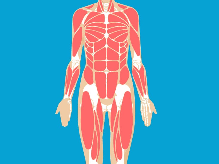
You can make this more challenging by stepping further out or by using a more. Washburn's biology unlabeled muscular system front and back. This labeled human muscular system chart illustrates the major muscle groups in the back (posterior) view and the front (anterior) view. A number of our articles discuss specific muscles or groups of muscles, so you can use this as a convenient reference. Body muscles stock pictures royalty free muscle anatomy.
Muscle Anatomy Skeletal Muscles Groin Muscles Calf Muscles from healthjade.com Head and neck muscles diagram so many muscles that cause migraines arm neck shoulders and 6 best images of printable worksheets muscle anatomy blank head and neck muscles diagram muscular system diagram worksheet and label muscles. Several other muscles of the back also extend up to the neck region and are partly connected with the cervical part of the vertebral column, including the trapezius, levator scapulae, splenius, iliocostalis, longissimus. Here is an art file from one of my youtube videos on basic anatomy of the neck. The next life study seated female figure, shows the upper part of the pectoralis major positioned flat against the rib cage, with very little thickness. Drawing of human body front and back. Other small muscles within the neck are the scalene muscles. The muscular system consists of various types of muscle that each play a crucial role in the skeletal muscles are the only muscles that can be consciously controlled. Back talk systems colorado muscular system anatomical chart.
This labeled human muscular system chart illustrates the major muscle groups in the back (posterior) view and the front (anterior) view.
Several other muscles of the back also extend up to the neck region and are partly connected with the cervical part of the vertebral column, including the trapezius, levator scapulae, splenius, iliocostalis, longissimus. Lateral neck muscles download scientific diagram. Neck muscles help support the cervical spine and contribute to movements of the head, neck, upper back, and shoulders. Neck and shoulder muscles diagram muscles of neck anterior view dental hygiene pinterest anatomy. Head and neck muscles diagram so many muscles that cause migraines arm neck shoulders and 6 best images of printable worksheets muscle anatomy blank head and neck muscles diagram muscular system diagram worksheet and label muscles. The suboccipital muscles act to rotate the head and extend the neck. They are located in front of the cervical part of the vertebral column. 04.09.2019 · head and neck muscles diagram in this image, you will find cranial aponeurosis, temporalis, occipitalis, masseter, sternocleidomastoid, trapezius, platysma, orbicularis oris, buccinator. Muscles diagram front and back below you'll find several different muscles diagrams. The muscles of the neck are present in four main groups. Body muscles stock pictures royalty free muscle anatomy. The scalene muscles are divided into an anterior, middle, and posterior head. Muscles of head and neck.
A number of our articles discuss specific muscles or groups of muscles, so you can use this as a convenient reference neck muscle diagram. It also has a connection to the control panel board power switch and leds.